
|
3D image rendering and animation of in vivo sample 3D confocal imaging of wounded mouse skin stained with 1% acridine orange in vivo. Dead cell nuclei over a muscle layer can be clearly seen. Cranfield C.G, P.M. Delaney, L.J. Bussau, R. G. King, and D.H. Barkla. (1996) Confocal Subsurface Microscopic Skin Imaging for Assessment of Wound Healing In Vivo. Proceedings of ASCEPT 3: 126. |
|
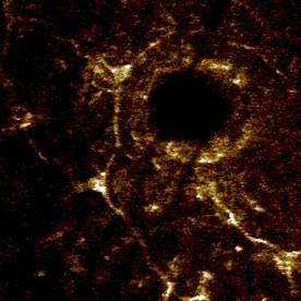
In vivo nerve imaging A neuron surrounding a hair follicle in hairless mouse skin in vivo. 0.1 ml of 100 mM 4-di-2-ASP was administered by intradermal injection. Image taken with an Optiscan F900e confocal microscope. Field of view = 250 µm |
|
In vivo blood vessel imaging In vivo image of blood vessels in hairless mouse skin following a 0.1ml intracardial injection of FITC-dextran (150kD). Image captured with an Optiscan F900e confocal microscope. Field of view =365 µm. |